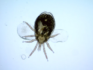
Answers Q 10: AMYLOIDOSIS
- Callithrix humilis
- Callithrix jacchus
- Callithrix nigriceps
- Callithrix commonus
 Which special stains are recommended for definitive diagnosis of the process affecting this common marmoset spleen?
- Gram stain
- Acid fast stain
- Congo red stain
- Periodic acid-Schiff stain
Which of the following other tissues are most likely to be affected by the same process?
- Brain
- Adrenal gland
- Skin
- Urinary bladder
Which of the following is the most likely cause?
- Chronic inflammation
- Dietary imbalance
- Space-occupying pulmonary mass
- Insomnia

- Callithrix humilis
- Callithrix jacchus
- Callithrix nigriceps
- Callithrix commonus

Which special stains are recommended for definitive diagnosis of the process affecting this common marmoset spleen?
Answers Q9 on Behavior

1) A nonhuman primate has been observed biting itself in the past, but has never caused any wounds. It has been treated for several months with twice daily diazepam (valium), with no convincing improvement. Recently, diazepam treatment was discontinued and treatment with fluoxetine (Prozac) was begun. Within a few days of this change, there was a noticeable increase in the amount of self-biting, and even some mild wounds found for the first time.
a. What are some possible causes for the increase in biting and wounds?
i. There are many possible external causes for any increase in an abnormal behavior, requiring a little behavioral detective work to identify. Did something change in the patient’s social structure, such as addition, removal, or even just relocation of an animal within the room? Has husbandry changed, either in the personnel the animal sees or how they act? Has there been a change in the experimentally that might impact the patient, such as disease progression, surgery, handling procedure, etc.? It is difficult to predict how changes such as these will affect animals, so an effort should always be made to identify potential contributors to an observed increase in an animal’s stress.
ii. Self-injurious behavior (SIB) can also be both cyclical, with better and worse periods not necessarily connected to treatment or external changes, and progressive, getting worse over time even in the face of effective treatment. In the above scenario, it is possible that the timing of the worsening behavior is purely coincidental and unrelated to the change in treatment. However…
iii. Many of the drug therapies used to treat anxiety and abnormal behaviors cause chemical dependence with use, and sudden discontinuation of these therapies is likely to trigger withdrawal symptoms regardless of whether the therapies were effective in the first place. These withdrawal symptoms will predictably increase anxiety and abnormal behaviors, so these drugs should always be weaned slowly, and a plan put in place prior to weaning of how to proceed if an animal’s behavior worsens during the process. Additionally, many therapies gradually become less effective with continuous use, requiring higher and higher doses or losing effectiveness completely. Benzodiazepines like diazepam pose both of these problems, losing effectiveness with time and causing serious withdrawal symptoms with sudden discontinuation. See #1 Janhsen et al. 2015, for a discussion of this problem in human medicine. In the scenario above, it is very likely that diazepam withdrawal is contributing to the increase in SIB.
b. Can we determine from this whether the fluoxetine is likely to be effective?
i. Fluoxetine has been shown to be effective within the first 4 weeks of treatment (#2 Fontenot et al. 2005), but it may take up to 3-4 weeks before the maximum effect is observed. Treatment with fluoxetine or other selective serotonin reuptake inhibitors (SSRIs) thus requires some commitment and a plan for determining effectiveness. It should also be noted that, as discussed above, these drugs should be expected to cause chemical dependency by the time their effectiveness can be determined, so almost always require slow weaning regardless of whether they are effective.
c. Other learning issues: As discussed in scenario 3, diazepam and other benzodiazepines may be effective in reducing anxiety associated with discrete triggers (cage transfer, cleaning, etc.), but the only systematic examination of their effectiveness in treating SIB found that they are just as likely to make matters worse as better (#3 Tiefenbacher et al. 2005). Thus, diazepam and other benzodiazepines should probably not be a first line treatment for SIB of unknown cause.

a. What is the likely etiology of this hair loss?
i. The well-defined borders and concentration on the face and head make this very likely to be barbering, the removal of hair from oneself or cage mates. The whiskers are often, though not always, removed as part of this behavior. A good description of this behavior can be found at: http://web.stanford.edu/~jeromeg/cgi-bin/Barbering.php
b. Is this similar to any human disorders?
i. This behavior is considered a model for human trichotillomania, a disorder in which people pull and often consume hair (#4 George et a. 2015)
c. Are there any effective treatments?
i. Treatment with N-acetylcysteine, an amino acid anti-oxidant, has proven effective for treatment of barbering and for a related disorder, ulcerative dermatitis, in mice (#5 Vieira)

3) An otherwise friendly
dog has been reported to be increasingly stressed around the time of cage
cleaning each day. It started with lots of barking and whining, but has
recently turned into cowering at the back of the cage. Yesterday during
cleaning, the dog was cowering and then suddenly lunged at the care taker, who
barely avoided being bitten.
a. What are some
potential treatments in this case?
i. Fearful reactions to husbandry or
other routine procedures may not be the norm, but they aren’t rare. A
good place to start in troubleshooting such cases is to examine the procedure
to see if anything may have changed and/or if it is being performed
correctly. For example in this case, is something causing the animal to
get wet during cleaning, such as a faulty hose nozzle or new, untrained
technician? Has there been a recent change in technician to someone who
may not be as gentle or trained in how to clean without causing stress?
You should ask questions such as these, but also know that in many cases there
may not be any single inciting cause. Fearful reactions may slowly
progress because they are self-reinforcing in some way – in this case picture a
dog who reacts to the novelty of cage cleaning in a new facility by reluctance
to move. By this reaction, the dog does not move away from the spray
during cleaning and gets wet. The dog then associates this unpleasant
experience with the cleaning, reacting the next day with more fear and more
reluctance to move. You can see how, over time and perhaps without the
technician even noticing a change, the dog’s fear increases to the point that
eventually it reacts with aggression.
ii. The most effective treatments for cases
like this are often changes in husbandry or behavioral management, rather than
drug therapies. For these to be effective, one must do a bit of
behavioral detective work, observing the animal and attempting to determine
exactly what is triggering its anxiety response. In the case above, let
us suppose you have determined that the dog’s fearful response is tied closely
to the hosing of its cage, and does not appear related to a particular care
person or other stimulus like an aggressive neighbor. In the many cases
where such a discrete, husbandry-related trigger can be identified, a simple
change in management practices may completely solve the problem. In this
case, for instance, it might be possible to let the dog out of its home cage
during cleaning, allowing it to roam up and down the aisle during the stressful
hosing. Not only would this break the cycle of the negative stimulus
(hosing) and the animal’s anxiety, it would also add a daily enrichment for the
animal that may decrease its general anxiety level. Of course, in many
cases a trigger can be identified but it isn’t possible to simply avoid by
changes in management practices. In these cases, positive reinforcement,
paired with trigger, may be used to counteract the anxious reaction. This
process is called desensitization. In the example above, it would start
by finding a stimulus that is very rewarding for the dog (treats, petting, a
certain toy, etc.), then giving that reward while slowly re-introducing the
hose. Desensitization is a slow process that requires informed planning
and a skilled trainer, but the results can be very rewarding.
b. Are there any drug
therapies that might be contraindicated?
i. In considering a case like this, where
it is clear that the animal’s reaction is related to anxiety, it may be
tempting to treat with a benzodiazepine like diazepam. Benzodiazepines
are anxiolytic and used frequently in human psychiatry and veterinary medicine
to treat acute onset anxiety, whether triggered by a known stimulus or
not. However, in addition to their anxiolytic properties, benzodiazepines
cause disinhibition, and this is a potentially dangerous problem in cases of
fear-related aggression like in the case above. In these cases,
benzodiazepines are likely to remove the animal’s natural inhibition against
biting a human, resulting in more aggression rather than less. So – in
cases of fear aggression, especially in a species like dogs where handlers may
be in direct contact with the animal, benzodiazepines should usually be
avoided. If there use is absolutely necessary, they must be used with
extreme caution.
c. Would your drug
choices differ if this was a macaque who was reported to “lose it” once a week
during cage change?
i. In contrast to our dog example,
benzodiazepines may be effective for treating acute anxiety in animals that
don’t or cannot (due to lack of contact with handlers) react with fear
aggression. Midazolam, a very fast onset, short-acting benzodiazepine, is
especially effective when given 5-10 minutes prior to a procedure that lasts
less than an hour. Also, due to its short half-life, this drug is less likely
to cause the chemical dependence that is known to occur with repeated use of
longer lasting benzodiazepines, especially if used in conjunction with a
procedure like cage change that usually only occurs weekly or
semi-weekly. While they may be effective in these cases, benzodiazepines
must still always be used with caution. One study has demonstrated that
in animals with abnormal behaviors like self-injurious behavior, benzodiazepine
treatment may actually make the behavior worse, likely due to the disinhibition
discussed above (Tiefenbacher S, Fahey MA, Rowlett JK, Meyer JS, Pouliot AL,
Jones BM, Novak MA. The efficacy of diazepam treatment for the management of
acute wounding episodes in captive rhesus macaques. Comp Med. 2005
Aug;55(4):387-92.)
4) You are called to
examine a 7 y/o male rhesus macaque who arrived in your facility at the age of
5 with frustratingly little detail its medical record up to that point.
Multiple attempts have been made to socially house this animal, but it doesn’t seem
to respond to social cues normally, which has led to fighting each time.
You find the animal awake and alert, but curled into a ball and rocking while
sucking its thumb. On closer inspection, you find lesions on the arms and
legs that look like the picture below.

a. From this information,
can you determine a likely part of this animal’s history prior to arriving in
your facility?
i. The inability to respond normally to
social cues, coupled with the curled posture and thumb sucking, are indicative
of an animal with an abnormal rearing history, likely peer or nursery
rearing. (Harlow HF, Mc Kinney WT. 1971. Nonhuman primates and psychoses.
J Autism Dev Disord 1:368–375; Rommeck I, Gottlieb DH, Strand SC, McCowan B.
2009. b. The effects of four nursery rearing strategies on infant behavioral
development in rhesus macaques (Macaca mulatta). Journal of the American
Association for Laboratory Animal Science, 48 (4): 395- 401.)
b. What treatments might
you consider in this case?
i. Abnormal rearing is known to be
associated with self-injury in nonhuman primates, and these wounds are typical
of those seen in cases of self-injurious behavior (SIB). (Novak MA, El-Mallah
SN, Menard MT. Use of the cross-translational model to study self-injurious
behavior in human and non-human primates. ILAR J. 2014;55:274–28)
Additionally, abnormal rearing affects a number of behavioral and biological
systems, and in the case of SIB, the most important of these is likely the
serotonin system of neurotransmission in the brain (Kinnally EL, Lyons LA, Abel
K, Mendoza SP, Capitanio JP. The Effects of Experience and Genotype on
Serotonin Transporter Gene Expression in Response to Maternal Separation in
Infant Rhesus Macaques. Genes Brain and Behavior. 2008;7(4):481–486.; Champoux
M, Bennett A, Shannon C, Higley JD, Lesch KP, SJ S. Serotonin transporter gene
polymorphism, differential early rearing, and behavior in rhesus monkey
neonates. Molecular Psychiatry. 2002;7(10):1058–1063.), and serotonin
transmission dysfunction has been implicated in self-injurious behavior in both
humans and nonhuman primates (Novak et al. 2014). Based on this, treatments
targeting serotonin dysfunction would be a good place to start for this
case.
ii. Tryptophan is an amino acid present in
normal food that is the precursor for serotonin, and has been demonstrated to
be effective in treating SIB (Weld
KP, Mench JA, Woodward RA, Bolesta MS, Suomi SJ, Higley JD. Effect of
tryptophan treatment on self-biting and central nervous system serotonin
metabolism in rhesus monkeys (Macaca mulatta) Neuropsychopharmacology. 1998;19:314–321).
iii. Selective serotonin reuptake
inhibitors have also been shown to be effective in treating SIB in nonhuman
primates, though these take longer to take effect and may cause chemical
dependence, requiring slow weaning when discontinuing the drug (Fontenot MB,
Padgett EE, 3rd, Dupuy AM, Lynch CR, De Petrillo PB, Higley JD. The effects of
fluoxetine and buspirone on self-injurious and stereotypic behavior in adult
male rhesus macaques. Comp Med. 2005;55:67–74.; Fontenot MB, Musso MW, McFatter
RM, Anderson GM. Dose-finding study of fluoxetine and venlafaxine for the
treatment of self-injurious and stereotypic behavior in rhesus macaques (Macaca
mulatta) J
Am Assoc Lab Anim Sci. 2009;48:176–184.).
c. Once you begin
treatment, how will you determine whether it is working?
i. When treating self-injurious or any
other type of abnormal behavior, the most common way to determine whether
treatment is working is to observe the animal’s behavior in a controlled
way. “Controlled” in this case means that the behaviors of interest are
well defined, that the observations are of sufficient length to be
representative of the animal’s full day, and that the measurement is actually
reflective of the underlying pathology. For instance, you may want to
observe how frequently the animal is biting itself, or how long in a 20 minute
period is spent pulling hair, etc. Whenever feasible, these observations
should be made using video recordings or neutral observers (e.g. not the
veterinarian associate with injections or the care person associated with
feeding). These measurements should be made prior to beginning treatment,
though in cases of acute injury waiting for baseline measurements may not be
possible. In these cases, or where direct observation of the behavior
isn’t feasible for some other reason, you may want to measure physical symptoms
of a behavioral problem. This has been done for alopecia resulting from
hair pulling
(Bellanca RU, Lee GH, Vogel K, Ahrens J, Kroeker R, Thom JP,
Worlein JM. A simple alopecia scoring system for use in colony management of
laboratory-housed primates. J Med Primatol. 2014 Jun;43(3):153-61. doi:
10.1111/jmp.12107. Epub 2014 Feb 27.).
Relevant to our example case,
measuring the severity and locations of wounds is an effective method of
measuring SIB and is correlated to directly observed behavior (Freeman ZTKC,
Rice KA, Adams RJ, Metcalf Pate KA, Hutchinson EK. Severity and distribution of
wounds in rhesus macaques (Macaca mulatta) correlate with observed
self-injurious behavior. J Am Assoc Lab Anim Sci. 2015 Sep;54(5):516-20.)
1: Janhsen K, Roser P, Hoffmann K. The problems of long-term treatment with benzodiazepines and related substances. Dtsch Arztebl Int. 2015 Jan 5;112(1-2):1-7. doi: 10.3238/arztebl.2015.0001. Review. PubMed PMID: 25613443; PubMed Central PMCID: PMC4318457.
2: Fontenot MB, Musso MW, McFatter RM, Anderson GM. Dose-finding study of fluoxetine and venlafaxine for the treatment of self-injurious and stereotypic behavior in rhesus macaques (Macaca mulatta). J Am Assoc Lab Anim Sci. 2009 Mar;48(2):176-84. PubMed PMID: 19383215; PubMed Central PMCID: PMC2679666.
3: Tiefenbacher S, Fahey MA, Rowlett JK, Meyer JS, Pouliot AL, Jones BM, Novak MA. The efficacy of diazepam treatment for the management of acute wounding episodes in captive rhesus macaques. Comp Med. 2005 Aug;55(4):387-92. PubMed PMID: 16158915.
4: George NM, Whitaker J, Vieira G, Geronimo JT, Bellinger DA, Fletcher CA, Garner JP. Antioxidant Therapies for Ulcerative Dermatitis: A Potential Model for Skin Picking Disorder. PLoS One. 2015 Jul 13;10(7):e0132092. doi: 10.1371/journal.pone.0132092. eCollection 2015. PubMed PMID: 26167859; PubMed Central PMCID: PMC4500395.
5: L. T. Vieira G. Preventing, treating and predicting barbering behavior in C57BL/6 mice. Ph.D., Purdue University. 2014. Available at: http://docs.lib.purdue.edu/dissertations/AAI3669652/
Answers Q8: Infections on research

Venturing into the immune system is not my area of expertise so forgive the simplification. Here is the background: for many years we had a problem with isolated pockets of pinworms or mites in localized areas of our mouse facilities. Of course these become much less localized when investigators collaborate and share mice... and indeed some pinworm species (notably Syphacia muris of rats and Aspiculuris tetraptera of mice) have been demonstrated to have eggs that persist for long periods in the environment. So when we had a problem that could relate to the immune system, the first thing we did was look for parasites. In this case we suspected that the culprit may have been pinworms or mites - why? because nematodes (and fur mites) switch the T helper cell response from Th1 to Th2 predominance, and the Th1/Th2 balance (where Th1 predominates) plays a key part in producing the EAE phenotype. Remember the Th1 cell primarily acts against intruders inside the cell (viruses and intracellular bacteria) whereas Th2 cells act against intruders outside the cell (bacteria and parasites).
Here is a direct quote from the abstract of Romagnani S. Ann Allergy Asthma Immunol. 2000 Jul;85(1):9-18; quiz 18, 21.
"Th1 cells, which produce interferon (IFN)-gamma, interleukin (IL)-2 and tumor necrosis factor (TNF)-beta, evoke cell-mediated immunity and phagocyte-dependent inflammation. Th2 cells, which produce IL-4, IL-5, IL-6, IL-9, IL-10, and IL-13, evoke strong antibody responses (including those of the IgE class) and eosinophil accumulation, but inhibit several functions of phagocytic cells... Th1-dominated responses are involved in the pathogenesis of organ-specific autoimmune disorders...Th2 responses are responsible for atopic disorders."
2. I have a mouse model investigating immune responses in our genetically engineered mouse model that has cloned T cells (limited action only to defined epitopes) - however the controls as well as the test animals are both reacting the same. Is it norovirus?
Norovirus has been a very popular mouse virus - even my investigators had heard about it! Like in the first question, the investigators were having a problem with an immune response, although in this case the mouse was responding when it shouldn't have. i.e. something was eliciting a response. The first thing to note was that the mice in question were immune deficient - their T cells couldn't respond to the usual panoply of organisms, so we could anticipate that they might be susceptible to an opportunist. The most common opportunists we see are MNV, Pasteurella pneumotropica and various other bacteria. MNV wasn't top of my list because at that time little was known about it except it had been reported to cause problems in mice deficient in the Jak–STAT1 pathway. Regardless, we tested the colony and found it was positive for both MNV and P. pneumotropica. We were busy discussing the next step (rederiving the mice) when the PI noted that he had a second colony of these mice in another building. We tested those mice and discovered that in this case they were positive for MNV, but negative for Pasteurella. Perfect! We suggested running a trial experiment on these mice to see if they would had the same aberrant response. As this was a lot easier than cleaning up the mice, the investigator agreed. Result: the experiment worked perfectly, proving MNV was not the culprit - (my bets were still on Pasteurella though).
3. I'm losing my mouse line ... it's a transgenic model I created and so far as I know there are no immune deficiencies. However I'm getting infertility, small litter sizes and unexpected deaths in young adult mice. Help!
This is a short one: this is a very common scenario for P pneumotropica in genetically engineered mice where the immune system has not been characterized (likely there is something that is not yet known, anyway). In fact it's so common that we automatically treat for it when we hear this story.
Answers Q7: Endoparasites
 Mite from a locally wild-caught big brown bat. I'm not an expert on bat mites, but it is likely this is the macronyssid mite Steatonyssus occidentalis (as described in Pearce and O'Shea, Journal of Parasitology 93(3):518-530. 2007) It bears obvious similarities to Mesostigmatid mites of rodents (see next) and blood sucking mites such as the Northern fowl mite (Ornithonyssus sylviarum) tropical fowl mite (O. bursa) and poultry red mite (Dermanyssus gallinae) of birds. These mites are all similar in appearance but spend variable amounts of time in the environment vs. on the host, so knowing the epidemiology is really useful for eradication purposes.
This one is Ornithonyssys bacoti - the tropical rat mite - though it was the subject of a mouse infestation so they are obviously not picky as to their food. They bite people readily too, and in parts of the world where tropical diseases are found, they can transmit some pretty nasty tropical diseases. Notice the beautiful little suckers on the ends of the legs - these disintegrate when they die so pictures of dead mites don't have them...

The next one was part of the same outbreak as the Ornithonyssus above. They both arrived as a result of a major renovation project in another part of the building. In fact these fast-moving mites that generally lay their eggs in the environment are generally found on wild rodents, but if you remove their wild hosts, they have no problems traveling long distances to find another host, such as your laboratory mouse colony. You notice that it's much more chitinized than the previous mites - in fact this was a preserved specimen but even in fresh specimens the chitinization is obvious and it's not so easy to see their blood meal inside. It's the spiny rat mite (Laelaps echidninus).

The next one should be much more familiar to you: it's one of those fur mites that many of us found out we had once we started PCR testing racks! Look for the body bulges between the legs - that tells you it is a Myobia or Radfordia. These 2 genera are almost identical, but if you look closely at the second pair of legs (don't get confused, the first pair are those stubby things pointing forwards, handily modified for hair-grasping) you can tell how many "claws" there are on the end. This one has 2, which makes it a Radfordia. And if you want to get picky, unequal claws mean it's a mouse Radfordia (R. affinis) as opposed to equal claws on the rat fur mite (R. ensifera)

This picture was actually taken to demonstrate the claws on digit 2, but it really just shows how hard it can be. Is this a single claw (Myobia)? or 2 unequal ones (Radfordia)? Your guess is as good as mine - however the action required is the same - determine the extent of the outbreak (hope your PI doesn't have thousands of cages, and dozens of collaborators), treat and retest. These days we check following treatment by PCR testing the racks, which have to be washed and cleaned of DNA of course, but that's another story.
 This one is the other fur mite - Myocoptes musculinus - notice the difference in body shape, the front legs actually look like legs, and this one has 2 pairs of huge back legs too (this is a male, they are less obvious in the female). These mites are a lot more mobile than the Myobia/Radfordia mites. However all these fur mites live entirely on the host, and even Myocoptes mites don't move more than a few inches off the host even if it dies.
Demodex musculi. I stole this picture from the internet because it's better than my pictures, but we too have actually found these on 'normal' mice using a deep skin scrape. In the past no-one really looked too hard for them but of course, PCR is showing that they are present, and they are being reported as a problem in immunocompromised mice, as described in the recent paper Smith et al: Comp Med. 2016;66(4):278-85 .

I hope you weren't fooled by this one - looks like plant material to me!
 This is from a rabbit - usually found in the ears but you can occasionally find them in a localized patch on the skin too (I don't know how often - I found it once!) Otodectes cynotis, the rabbit ear mite

This delightful creature isn't a parasite at all - it's a book louse or Psocid. However you may be unfortunate enough to find it on the feed bags or even in the feed. If you do it's probably a sign that the food bag, or feed was stored at or subjected to high humidity.

This one was found in a fecal float from a mouse. Definitely not a rodent fur mite - but what exactly, I'm not sure. Looks like a mite of some sort, probably arrived in the feed and still recognizable after being passed through the digestive tract of the mouse.
How did you do?
Goeldi’s monkeys – Callimico goeldii (callimico is Latin for beautiful little monkey) are related to marmosets. They are reclusive inhabitants of the understory of the Amazon rainforest. They have been observed living in groups of up to 11 monkeys usually with only one female per group. Unlike marmosets (which usually have twins) or other NHP, they often have 2 single infants per year.


Answers Q6: Red Cells
 |
| Image from Wikimedia |

What is a Heinz body?
Heinz bodies are RBC inclusions consisting of denatured hemoglobin. Heinz bodies are usually indicative of oxidant-induced hemolytic anemia. However In marmosets up to 5% of RBC containing Heinz bodies are considered normal.
Are they likely to be a common finding in Goeldi’s monkeys?

Goeldi’s monkeys are closely related to marmosets (they are sometimes called Goeldi's marmosets), so yes. Small numbers can also be seen in normal cats.
Bonus: what is the genus and species of a Goeldi’s monkey?
Callimico goeldii. Family callitrichidae
What pathological process do Heinz bodies normally indicate?
In humans, Heinz bodies are indicative of unstable hemoglobin or splenic dysfunction. They were first described in 1890 in association with hemolytic anemia by Robert Heinz, a german physician. Oxidation of sulfhydryl groups on Hb results in precipitation of Hb and the formation of the small intracellular inclusions called Heinz bodies
Give an example of an inherited disorder that results in this pathology
Inherited G6PD deficiency or NADPH deficiency can also result in oxidative damage to RBC and Heinz bodies.
Apart from inherited disorders, what other conditions can result in this pathology in animals?
In cats, dogs and NHP, they are usually associated with consumption of acetaminophen, garlic or onions. In cats, ingestion of dog food containing propylene glycol. They can also be seen in chronic liver disease, or splenic malfunction.
What stain is often used to highlight Heinz bodies?
They are not easily seen with Romanovsky stains such as Giemsa but are well demonstrated with supravital stains such as new methylene blue.
Marmosets also often have Howell Jolly bodies in their blood. What is a Howell Jolly body?

Howell Jolly bodies are small nuclear fragments that were not extruded when the RBC left the bone marrow. They present as a perfectly round blue inclusion.
The presence of a large proportion of Howell Jolly bodies usually indicates what pathology?
They are often a sign of regenerative anemia. They are normally removed by the sinusoidal spleen so increased numbers may be seen if the spleen is not functioning – also normal cats and horses may have low numbers perhaps because their spleen is non sinusoidal.
References and more information:
https://en.wikipedia.org/wiki/Heinz_body

Which malaria spp. are used to study blood stage malaria in the rhesus host?
Falciparum ~= P coatneyi (and P fragile) from cynomolgus monkeys in a rhesus model. Has many characteristics of human falciparum malaria, inc. multiple ring forms in one RBC, applique forms, RBC rosettes, sequestration in certain organ vascular beds by adhering to endothelial cells via CD36 and severe sequelae including cerebral malaria and severe anemia.

 Which NWM model (malaria spp, NWM spp) is the only one to reliably produce the dormant liver stage of malaria?

Answers Q5: Malaria Models
 |
| From Autino et al. Mediterranean j of hematology and infectious diseases Oct. 2012 |
Background
Malaria is the most common parasite of humans with 100’s of millions (!) of clinical cases annually
4 commonest human species are P falciparum, P. vivax, P. malariae and P. ovale. RBC disorders such as sickle cell disease, ovalocytosis and thalassemias conveying some resistance to malaria may have evolved as a result of selection pressure exerted by malaria
What are the 2 hosts required for potentiation of malaria infections? (be specific)
All require 2 hosts, a female anopheles mosquito and a primate. ( if a Q says be specific, its a clue - don't forget to mention it's the female mosquito).
In general, can old world primates (OWM) be infected with human malaria spp. ? Can new world primates (NWM)?
OWM (with one exception) cannot be infected with human malaria spp. (nb. the reverse is possible: several OWM malarial spp. can infect humans). NWM Aotus and Saimiri can be infected with human malaria spp. Thus the OWM models are primarily OWM malaria spp. transferred to a new OWM genus or spp, and the NWM models are primarily human malaria spp.
In general what procedure is necessary to establish a malaria model into a new NHP family?
Splenectomy. * Bonus: why is the spleen removed?
What is responsible for the recurrent fevers characteristic of clinical signs of malaria?
Recurrent cycles of multiplication in RBC result in destruction of the RBC with release of merozoites and waste products. The waste products (hemozoin) and toxins (glycerol phosphatidyl inositol) cause fever and clinical signs that coincide with each cycle of replication.
In falciparum malaria, list the 3 different types of clinicopathology that can result in severe morbidity or death.
1. Cerebral malaria with multiple organ failure,
2. Severe anemia due to destruction of uninfected RBC by the spleen,
3. Hyperparasitism due to cycles of increasing blood multiplication uncontrolled by immunity.
 |
| From Wells et al. Cell. March 2010 p.145. Showing a schizont (top) and hypnozoite (bottom) |
Which human malaria spp. have a dormant stage in the liver, what are the dormant malaria forms called, and what is reactivation of dormant malaria called?
Vivax and ovale (not falciparum and malariae) can go dormant in the liver, the dormant forms are called hypnozoites. They can reemerge even years later, causing a “relapse."
Which human malaria spp. can cause severe glomerulonephrosis? Which causes ARDS?
Malariae, although often referred to as ‘benign,’ nevertheless can cause severe renal disease
Vivax malaria can cause severe pathology due to ARDS anemia and splenic rupture

OWM models infected with other NHP malaria spp are generally used to study the blood stage because the larger NHP size allows for both blood sampling and bone marrow samples. OWM malaria species including P coatneyi and fragile (from cynos), P cynomolgi, P knowlesi (from cynos and pigtails) and P inui are all used in rhesus. Rhesus models are also used to study the malaria of pregnancy.
Which malaria spp is considered to be the OWM analog of falciparum? Vivax? Malariae?
Which is the most prevalent natural malaria spp in OWM?
Vivax~= P cynomolgi (despite the name is found in several macaque spp from S Asia). It can be . mistaken microscopically for P vivax. Characteristics include Schuffners stippling seen in infected RBCs when stained with Giemsa – these morphologically are caveolae-vesicle complexes. The model also produces the hypnozoites in the liver with relapses like vivax.
Malariae~= P inui. It is the most prevalent natural OWM malaria in SE Asia.
Characterized by extended periods of development in both mosquito and primate and can cause chronic infections (years) with renal sequelae (glomerulonephrosis) like in humans. Can be zoonotic.

What kind of malaria studies are NWM used for and why?
Drugs, drug resistance, vaccines and acquired immunity. Because NWM can be infected with human malaria spp.
NWM used are primarily Aotus spp (nancymaae and vociferans are the most readily available to investigators). (trivergatus AKA griseimembra in home countries only) and Saimiri spp (sciureus b. boliviensis although boliviensis limited availability)
 Which NWM model (malaria spp, NWM spp) is the only one to reliably produce the dormant liver stage of malaria?
P vivax (Brazil VII strain) in Aotus spp.
Name the 2 spp of NWM malaria that are thought to be anthropozoonoses. Which is most common in the wild?
P brasilianum infects many NWM spp. and is fairly ubiquitous in the wild.
P simium (only found in Alouetta and Brachyteles in S Brazil) is morphologically identical to vivax in humans.
How is experimental malaria treated in NWM? Is it curative?
Yes, curative.
Simian malaria is sensitive to chloroquine 10mg/kg daily for 3d.
Relapsing malaria - primaquine sid for 7d.
Resistant malaria – mefloquine or CoArtem.
List the 2 types of model generally used for vaccine trials

Sporozoite challenges in OWM with simian parasites spp.
Blood stage (ring stage of RBC infection) challenges in NWM
* Bonus: The spleen is removed to facilitate adaptation of malaria to a new spp. (Once adapted, the malaria can be transferred to a less susceptible or spleen-intact model).
The spleen removes damaged red blood cells from the bloodstream by phagocytosis, and initiates innate and adaptive immunity. Splenectomy results in a greatly diminished frequency of memory B cells.[9] and increased disease susceptibility. In malaria the role of the spleen appears to be removal of younger ring-stage parasites from infected red blood cells without destruction of the RBC. Older >24h parasitized RBC adhere to vascular endothelium and thus are protected from splenic removal.
Reference: [The Blue Book ]Nonhuman primate models in biomedical research: (Elsevier 2012) Chapter 5 "Nonhuman primate models for human malaria research" by Mary Galinski and John Barnwell.
In which CITES appendix are these (wild) rodents listed?
What are the 2 species, from where did they get their name, and which of these species is bred in captivity?
Which features of their normal ecology affect our husbandry with respect to caging, cage furniture/enrichments, nutrition and group housing?
Answers Q4: Chinchillas
 |
| Chinchilla lanigera -wild type |
Why were these rodents hunted almost to extinction by the early 20th century?
- Chinchillas are native to the Andes in S America and were hunted for their dense, soft fur (up to 75 individual hairs per pore).
- They were part of the international fur trade as early as the 16th century, and as a result by the early part of 20th century they were almost extinct.
- Both species are listed in CITES appendix I (the most protected) – first listed in 1975.
https://cites.org/sites/default/files/eng/cop/09/prop/E09-Prop-09_Chinchilla.PDF
Why are they no longer under pressure from hunting in the wild?
- Captive breeding has produced larger chinchillas with much improved colors (see below compared with top picture).
 |
| Chinchilla lanigera - product of captive breeding |
- Chinchilla brevicaudata (ie short tail) and Chinchilla lanigera (literally - bearing woolen coat). The name Chinchilla comes from the Chincha peoples of the Andes and means ‘little Chincha’. The species bred in captivity is Chinchilla lanigera (AKA long tail, coastal or lesser chinchilla). C. brevicaudata is also known as the Bolivian, Peruvian or Royal chinchilla.
- Their skin produces abundant lipid and unless they have frequent (several times a week) dust baths their coat becomes greasy and lank. Dust baths are available as a mix of silver sand and fullers earth, volcanic ash or even diatomaceous earth. Talc should be avoided as it has been shown to cause pulmonary granulomas.
- They are shy and easily stressed – and crepuscular. Their cages should have a hiding place.
- They are very good at escaping – cages for other species need to be modified to remove gaps that their skull can go through.
- They are monogamous, females have a vaginal closure membrane and are seasonally polyoestrus q. 30-50d (but may breed year round in captivity).
- Females are larger than the males and prone to aggression post-breeding so the male needs a place to escape
- Kits are precocious born with hair and eyes open and eating solids within 7 days. (What other species do you know that are precocious?)
- Females have a urethral papilla that resembles a penis – anogenital distance is the best method for sex determination
What 2 main research areas are they commonly used for and why? Research with these rodents has substantially contributed to our knowledge of the pathogenesis treatment and prevention of which important human bacterial disease?
 |
| Organ of Corti from Bohne et al, Hear.Res.2017 Feb p158- |
- Infectious disease, particularly those causing otitis, with major contributions in the study of pneumococcal otitis caused by Streptococcus pneumoniae.
- Acoustic and acoustic toxicity studies because of a large tympanic bulla, easy access to the middle/inner ear and lack of intercurrent middle ear infections. Additionally, their hearing range is similar to those of humans (20 Hz – 30 kHz cf. humans 2Hz-20kHz). Other rodents can have hearing up to 100 kHz with peak sensitivity at frequencies at or above the upper limit of human hearing. The structure of their middle and inner ear and eustachian tube is also similar to those of pediatric humans.
- They were genetically sequenced in 2012 and there is a chinchilla research resource database http://crrd.mcw.edu/
Other information from Chinchilla. (2008, August 23). New World Encyclopedia, . Retrieved 15:40, February 20, 2017 http://www.newworldencyclopedia.org/entry/Chinchilla

 Their peripheral blood smears contains a unique estradiol dependent cell. What is its name and what kind of immune activity does it have?


Answers Q3: Hairless Guinea Pig
What is the genus and species of this animal?

Cavia porcellus
There are 2 types of this beasty without hair – one is born without hair and grows sparse hair mostly on the muzzle and feet (the “skinny” pig), and the other is born fully furred and begins to lose its its fur at 5 days of age becoming completely bald by 2m (the Baldwin pig)
What is the genotype of the commercially available strain (skinny)
Crl:HA-Hrhr
(From this you should now know that it is outbred, and has a (recessive) hairless mutation on the 'hair growth associated' gene that encodes for a protein involved with hair growth. Bonus: what does the HA stand for?
Does its hairlessness make it more vulnerable to injuries and infections?
Yes because normally the hair provides a barrier to damage, particularly on the feet.
What is its immune status?
Euthymic and immune competent
Bonus: do you know the genotype and immune status of the nude mouse, hairless mouse and nude rat?
 Their peripheral blood smears contains a unique estradiol dependent cell. What is its name and what kind of immune activity does it have?
Kurloff cell: natural killer cell activity. Note the large granular magenta inclusion body.

What is notable about guinea pig neutrophils?
They are known as heterophils due to the acidophilic or eosinophilic inclusion bodies in their cytoplasm. Below is a picture of a heterophil (right) compared with an eosinophil (left).

What is a common cause of bumblefoot found in poorly maintained or inappropriate caging?
Staphylococcus aureus
picture from www.guinealynx.info/
Unlike in mice and rats, guinea pigs are very susceptible to clinical bacterial infections. List 3 that are the most likely to be found in research colonies.
Bordetella bronchiseptica
Pneumonia, conjunctivitis, otitis media, abortions and stillbirths. Rabbits can be asymptomatic carriers – a reason for not housing rabbits and guinea pigs in the same airspace.
Streptococcus pneumoniae
Can be subclinical carriers in nares: environmental stressors can precipitate pneumonia and middle ear infection.
Streptococcus equi subsp. Zooepidemicus.
Can be latent in the nares: disease precipitated by oral cavity abrasions (for example from overgrown molars) and causes suppurative lymphadenitis (guinea pig “mumps”) often unilateral.
Q: Which of these is a zoonosis?
A: S pneumoniae.
(S equi zooepidemicus has been isolated from human throat swab, though. Humans have their own Bordetellas - B. pertussis or B. parapertussis)
See also the Blue Book 'Laboratory Animal Medicine' pp. 247 on.

Thierry Yandza et al in Journal of surgical research 2012
Answers Q2: Transplantation

Q: What are these 2 approaches to gut transplantation called?
A = heterotopic transplantation (In medicine, "heterotopia" refers to presence of a particular tissue type at a non-physiological site, but usually co-existing with original tissue in its correct anatomical location. In other words, it implies ectopic tissue, in addition to retention of the original tissue type). {definition from Wiki}
B= orthotopic transplantation. ( from the Greek - “in the normal place” ie instead of the original tissue)
Q: What is another name for “A” based on the people who first described it?
Thierry –Vella Loop
Q: List reasons that pigs are considered the gold standard for these surgical models, rather than the rat.
- Pig and hu ( and dogs and NHP) have all -glandular stomachs (mo and ra have squamous and glandular portions)
- Pig and hu have similar small intestine/body weight ration of 0.1 vs 0.16 in rodents
- Pigs have the potential for inter-species transplants
- Pig and hu (and mice) have finger-like villi vs leaf-like in rats
- Pig and hu colons have sacculations and muscular bands (tenia) resulting in similar transit times vs rodents do not
- Pigs and hu both have some fermentation in the large gut and have similar microbiota whereas 85% of rodent gut organisms are absent in humans
Useful resources include
Amanda Zeigler et al in cellular and molecular gastrointestinology and hepatology 2016 and
Answers Q1: Voles
Voles: fun facts
Subgenus Microtus
2 most widely used in research:
M pennsylvanicus (Eastern meadow vole)
M ochrogaster (prairie vole)
(pictured are prairie voles)
Widely distributed in N America
They eat mainly grasses which are high fiber low calorie. Why does this matter?
- We feed them rabbit food not rodent food
- On high fiber diets cellulolytic bacteria responsible for fermentation have been detected in the esophageal sac (of the stomach) resulting in a higher pH and VFA’s (like ruminants) than is normally seen in the stomach
- They are acclimated to eating often - they can get hypoglycemic in as little as 6hr without food
- They don’t do well in low temperatures (in contrast, mice can survive sub freezing temperatures by huddling and brown fat thermogenesis)
What is their normal habitat?
- Many species of voles are burrowing animals (in fact one species of vole, Pitymys, is somewhat adapted to underground (semifossorial) life like the naked mole rat (BTW do you know the genus and species of the naked mole rat?). Q: what are those adaptations do you think? Hence a solid bottom cage with lots of bedding is appropriate.
- Meadow voles are prone to stress induced seizures
Do they breed in captivity?
- Yes – but they are especially prone to cannibalism if disturbed in the first week after parturition and breeding cages should not be completely cleaned so as to maintain normal reproductive cycles.
- M ochrogaster are monogamous with the male assisting in parenting (Q: what other rodent species are monogamous?). In contrast M pennsylvanicus is promiscuous and sexes have separate nests.
- Most Microtus species mature early (can mate at 2-3 weeks) are very prolific with up to 17 litters a year possible.
Why are they used in biomedical research?
- Meadow voles have been used in nutritional studies and as bioassay animals for digestibility, protein content and toxicity.
- Prairie voles have been used extensively to study the physiology of the vomeronasal organ and how chemosensory cues affect behavior.
- Our prairie voles will be used to study distribution and binding to vasopressin of radioactive tracers in the brain.
What infections do they get?
- Good question. Many infections that rodents get in the wild have also been detected in voles. Eg hantaviruses, cowpox virus, Leptospirosis, T gondii, Echinococcus. Mites echinolaelaps and Psorergates, and blood parasites Babesia microti and Hemobartonella microti, plus others. A list of viruses such as we see for mice is not available.
No comments:
Post a Comment